Energy for Health, Vol. 2, 2008
Theofanous, N.°, Mathios, N.°, Grigoreas, P.°, Kordiolis, N.* Skarlatos, I.^, Pedrini, L.”.
° Laser&Optics Group, Department of Informatics, University of Athens, Greece.
* Department of Neurosurgery, Saint-Savvas Hospital, Ampelokipi, Athens, Greece.
^ Department of Radiotherapy, Saint-Savvas Hospital, Ampelokipi, Athens, Greece.
“ Department of Radiotherapy, IOV-IRCCS Padova, Italy.
Abstract
The conditions and characteristics of thermal / temperature response of a bulk PTC-type thermal sensor head (thermistor) irradiated by a CO2 laser beam are presented. Unmodulated (CW) and square-wave intensity modulated beam are compared. The results are evaluated by considering the sensor head as a physical mass and as simulating material for biological tissue of medical interest. it is thus derived that the intensity modulation, despite a slight lowering of the achievable temperature level, offers the advantages of improved temperature-to-optical power linearity, suppression of the corresponding saturation effects, and capability for temperature modulation.Introduction
It is well known that numerous interactions of a laser beam with biological tissue, for medical treatment, are mainly based on beam-induced thermal processes. Accordingly, the corresponding temperature ϑ and its rise created in the irradiated region and/or its adjacent layers play an important role for the achievement of the desired result (17). Indeed, the mean steady-state value of the tissue temperature, particularly when attained through absorption processes, determines the kind of the finally occuring effect which, for instance, may be coagulation or hemostasis if 60° c < ϑ < 100° c, cell water evaporation at 100°c, and pyrolysis or carbonization of the tissue at even higher temperatures (1-4. 8).
The above tissue temperature and the extent of its corresponding thermal effects are strongly influenced by the interaction time and the temporal form of the laser pulses directed to the tissue (1-3, 9). This fact implies that the timing of tissue temperature and its variations provoked by a repetitive/pulsed or a modulated laser beam may have significant influence on the effects in question. Unfortunately, as yet, in the field of clinical applications, modeling studies, interaction for of beam-tissue determining optimal treatment modalities exist only for the simple cases of unmodulated continuouswave (cw) or one-pulse laser beams with the real tissue roughly simulated by a single plastic material such as, e.g., swollen gelatin, ester methacrylates, or other (4, 6, 7, 10). By contrast, in the cases of laser beams with more complicated timing, such as the multiple pulses or intensity-modulated (iM) beams, no reliable modeling is in effect possible and therefore no trustworthy prediction of the corresponding tissue temperature can be made. This difficulty is enhanced by considering the eterogeneity of almost all living tissues (1-3).
In consequence, the implantation in vivo of an appropriate thermal sensor head (TSH) near to the irradiated tissue and the corresponding continuous monitoring of the actual temperature frequently constitutes the most reliable method for supervising laser-tissue interactions in the presence of thermal effects, particularly with variable/modulated laser light. lt is clear that, under these conditions, the thermal behaviour of the TSH mass itself (characterized by parameters such as its heat capacity, time of response, etc) play an important role in the correct evaluation and interpretation of thermometric results during the laser treatment. Moreover, in several types of thermal sensors, the thermally sensible medium is given the form of a relatively bulky head or it is encapsulated in such a head made by plastic material of dark colour, which is often the case of customary semiconductive thermistors (11-13). in such cases, the material of the thermal head can possibly simulate successfully enough a biological tissue and therefore the experimental study of temperature variations of such a head submitted to direct irradiation by a laser beam could provide a reliable prediction of the features of thermal interaction between this beam and the simulated tissue. This investigation would be even more useful in the case of modulated laser light, for which modeling data are absent or unreliable.
A first attempt in this direction has been made recently by Theofanous (14) using an electro-optically modulated Krypton laser beam incident on a thermistor head. However, in this trial a complicated set-up of external electro-optic modulation with sinusoidal format, which is rather unusual, was employed and moreover the spectral output of the Kr laser (red lines) did not match well the spectral absorption of the thermal head, which was composed of black-coloured material. Therefore, we have undertaken a similar study using a CO2 laser beam attacking a bulk-type PTG thermistor head, the black bakelite—like material of which can simulate acceptably dark biological tissues in thermal interaction with a CO2 laser beam.
In this paper, after a brief consideration on the features of heat deposition in tissues under modulated laser beam, we present the conditions and results of the above experimental investigation with the CO2 laser employed under stepwise cw regime and under square-wave intensity modulation. Results on the transient and steady-state temperature response are given for various modulation frequencies.
Laser-Induced Temperature and Timing Control
The value of temperature induced by a laser beam on an irradiated mass (thermal sensor head or simulated biological tissue) depends on the transfer of energy, which follows the general scheme depicted in figure 1, with the final interaction effect resulting from the combined action of reflection, absorption, and scattering of the laser light (4). among these phenomena that of absorption is the principally responsible for the elevation of temperature.
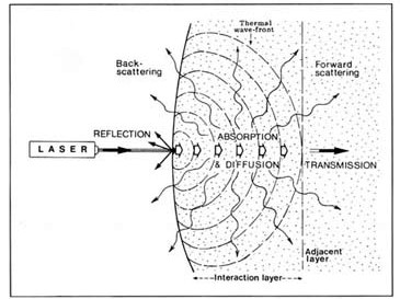
Figure 1
Typical scheme of thermal interaction between a laser beam and a sensible material (thermal sensor mass or biological tissue). The material in question is assumed homogeneous, so as the thermal wave-front propagates uniformly throughout the materials.
Generally speaking, the temperature of the irradiated mass is determined or affected either by material factors, which are related to the target material, or by beamrelated factors, characteristics of the laser beam itself. among the material factors, the more important are the absorption coefficient, the surface temperature, and the thermal conductivity of the material under irradiation (15). Note that thermal conductivity determines the amount of heat transmitted (undesirably) to the adjacent layers of an attacked tissue and therefore plays a significant role in surgical operations (16). on the other hand, among the beam-related factors, the more critical one is the timing control of the beam and it has been clearly ascertained that the steady-state temperature of an irradiated tissue decidedly depends on the ratio of the laser-tissue interaction time to the thermal diffusion time τa of the attacked tissular material (5-9, 17,18).
Unfortunately, the theoretical study of the above influence of timing control, has not but small practical use as no reliable information on absorption or scattering parameters of tissues is currently available and the corresponding transport equations require elaborate mathematical procedures (19). Therefore, as yet, investigations of this kind have been performed mainly in the experimental field and moreover – for simplicity – only with interrupted cw and spontaneously / autonomously pulsed or Q-switched lasers.
By contrast, to the authors’ knowledge, investigations using intensity modulation (iM) of the laser light, for external control and timing of its optical power or intensity, are lacking in the literature. This light modulation offers a number of advantages (capability of freely adjusting the period and height of laser pulses, possibility of commanding the beam, etc ) and therefore has been considered in the present work.
Experimental System and Procedures
The experimental set-up used in the present work is depicted schematically in figure 2. The beam of a co2 laser source, after passing through a series of Si attenuation filters (which control the optical power P of the beam emerging from their set), attacks the front surface of the thermal sensor head. in the particular experimentation, as co2 laser source the lM-4 model of edinburgh instruments co has been used which emits at a stabilized wavelenght of 10.6 µn, with internal water cooling, and offers the possibility to modulate its intensity – if it is desired – by means of a square-wave generator controlling straight- forwardly the current of the laser source. The amplitude of the output of the square-wave generator and the frequency of its square waveform, which is equal to the modulation frequency of the laser beam, were monitored by means of an oscilloscope continuously connected to the generator.
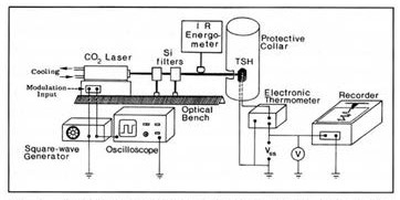
Figure 2
Arrangement of the experimental set-up employed in the present work for measurements on the thermal interaction between CO2 laser and the materials of the thermal sensor head (TSH).
The nominal optical power emitted from the above laser source, in cw operation, was of the order of 350 mw, for a temperature of the device equal to14° c ± o.5° c which has been maintained constant throughout the experiment by means of the internal closed cooling system. Nevertheless, for the purposes of our experimentation, the (mean) laser power incident upon the thermal sensor was less than the above cited value, depending on the particular combination of the attenuating Si neutral-filter plates and the frequency of intensity modulation. Therefore, the incident mean laser power was each time measured by means of an appropriate i.r. radiometer/energometer inserted as it is shown in figure 2.
On the other hand, the thermal head, used as thermally sensitive target simulating dark biological tissue and at the same time as temperature measuring element, was a bulk PTc semiconductor thermistor of the KTY 10 type of Siemens co. This thermistor was connected as thermally variable resistor rϑ in the electric bridge of the electronic temperature measuring circuit shown in figure 3. This circuit has been designed and constructed specifically for the needs of the present experimentation on the basis of design suggestions of related technical literature (11-13). we have thus succeeded in implementing an electronic thermometer which is highly sensitive and speedy in response (13) with a linearity largely enhanced by the electronic circuitry of the stage, particularly for the range from o to 100°c where a technical sensitivity of about 14 mv/°c has been achieved. Note that, as seen in figure 2, the output voltage vss of the electronic thermometer in question is continuously monitored by means of a digital voltmeter and recorded by a chart recorder.
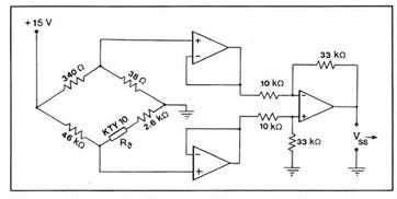
Figure 3
Schematic diagram of the electronic thermometer circuit used for the KTY10-type thermal head TSH, A11 operational amplifier are of the LM 741 type.
lastly, the thermal sensor head TSH was fixed on an adjustable micropositioner which was sheltered as shown in a large cylindrical housing made of a white cardboard sheet. This housing was intended to keep the temperature of the thermal head unaffected by any air currents invading the experimentation space. also, the white internal walls of the housing reemit most of the thermal radiation emitted by the thermal head while being under laser heating; in this way attempted we simulating the conditions existing in living tissues, in which – as already seen – a part of the laser energy emitted (by transmission of diffusion) from the targeted tissue region is back-scattered or rediffused by the surrounding tissue layers (fig. 1).
Measurements and Results
Preliminarily, we have graduated our electronic thermometer by extracting accurately the graduation curve vss versus ϑ, where ϑ denotes the reading in 0c of a high-precision centigrade mercurial thermometer, immersed together with our thermal sensor, head in water progressively heated up to 100°c. This curve has shown that our thermometer possesses a technical sensitivity of 13.8 mv/0c which is satisfectorily precise and constant over the 0 to 70° c range and deviates slightly for higher temperatures.
first of all, we have determined the thermooptical step response of the thermal head, i.e., the time variation of its instantaneous temperature ϑ just after the application of a stepwise cw laser “pulse”, such as the one shown by the dashed line of figure 4. The corresponding step response curve, ϑ versus t, where t denotes time, has been extracted and represented straightforwardly on the chart recorder for various levels of the laser power P incident upon the thermal head. one representative curve of this kind, obtained for an optical power P equal to 100 mw (step value) is depicted in figure 4. from these step response curves, the mean value of the thermal time constant (rise time) τr of the thermal head, defined as the time period needed for a rise of temperature from the 10% up to 90% of its final steadystate value, has been determined and found to be of the order of 10 min.
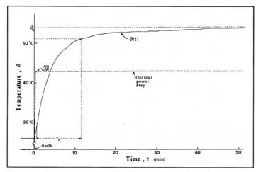
Figure 4
A representative transition and steady-state response curve ϑ(t), of the temperature ϑ of the attacked sensible materials (TSH) when an optical power step of 100 mW is incident upon it. ϑs denotes the steady-state of ϑ and t, the thermal time constant (rise time) of the materials mass under consideration.
from the same set of step response curves we have determined the dependence of the steady-state temperature rise Δϑs, produced by the beam on the thermal head, as a function of the cw-level power P of the step-shaped laser “pulse” (unmodulated) arriving at the sensor head. The so obtained Δϑs versus P curve is depicted in figure 5, along with the maximum-error margins associated to each individual determination of Δϑs.
Next, passing to the intensity-modulation regime, we have applied to the laser beam, a full square-wave modulation by driving the corresponding input of the laser source with a train, of square-wave voltage pulses based or, a level of 0 v and with an amplitude of 11 v. according to the manufacturer’s manual, an intensity modulated (iM) laser beam was then obtained; the modulation, depth was 100% and the modulation frequency fm, was equal to that of the square-wave voltage pulses (fig. 6). obviously, under this modulation, the mean power P of the laser pulses will be equal to the half of their maximum value (amplitude) Pm. Under these conditions, it has been ascentained experimentally that, in any case, the time temperature response of the thermal head to the above squarewave iM modulated beam has the form represented in figure 6a. as we observe from this figure, the temperature of the so irradiated thermal head, after an initial transient increase of undulating form, attains a varying steady state in which it oscillates, almost sinusoidally, about a constant steady-state mean (dc) value ϑs with an also constant peak-to-peal (ac) amplitude ϑo. To describe quantitatively the situation, it a convenient to use as principal parameter the ratio m = ϑo / Δϑs, which we shall call the temperature modulation depth.
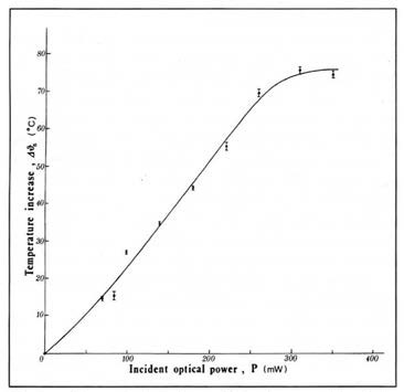
Figure 5
Measured dependence of the increase Δϑs of the steady-state temperature of the thermal head material upon the level of the (stepwise) incident optical power P of the laser beam unmodulated. The initial temperature of the thermal head with equal to 26°C.
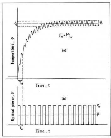
Figure 6
Shape of the temperature-to-power time response (a) of the TSH material when subjected to a laser beam intensity to a laser beam intensity modulated in the square-wave form shown in (b). For the various notations see the text.
We have monitored and recorded the time variations of the instantaneous temperature ϑ of our KTY-10 sensor head irradiated, in a stepwise manner, by various mean incident powers P of the laser beam being square-wave modulated at various modulation frequencies (repetition rates) fm ranging from 0 Hz (i.et unmodulated) up to about 0.8 Hz. from the monitored data we have derived that the dependence of the steady-state temperature increase Δϑs on the mean power P of the laser beam, for various modulation frequencies fm, is described by the curves given in figure 7.
In addition, from those of the above recordings which concern a standard incident mean power of 100 mw we have extracted the corresponding variations of the amplitude ϑo of temperature oscillations and the temperature modulation depth m = ϑo / Δϑs with modulation, frequency/repetition rate fm. These variations are plotted in figure 8 by the curves a and b, respectively, along with the corresponding depend¬ence of the temperature increase Δϑs on the frequency fm (curve c).
Lastly, from the same recordings it has been ascertained that the mean time constant (thermal rise time) of the sensor head does not undergo but a slight decrease with increasing fm and continue to be of the order of 10 min.
Analysis and Discussion of Results
first of all, as seen from figure 5, in the case of an unmodulated laser beam the increase Δϑs in the steady-state temperature of the irradiated material (which, in our case, is simulated by the medium of the thermal sensor head) proved to be an increasing but non-linear function of the step-wise incident optical power P of the laser light stream. Moreover, as seen from the figure, at higher optical powers the nonlinearity in question turns particularly marked and the Δϑs – P curve enters a characteristic saturation region and tends to become horizontal; hence no additional temperature increase is further possible, no matter how much we further increase the incident laser power. This saturation effect should be attributed to thermal compensation phenomena, such as the balancing between the light energy absorbed by the thermally sensible mass and the amount of energy re-emitted by thermal radiation, while taking into account the dispersion of energy due to the scattering processes quoted in the 2nd section (fig.1).
anyway, in practice the foregoing nonlinearity and saturation features of the Δϑs – P curve may be the cause of non-negligible complications, particularly when the laser beam system is used for treatment or biomedical purposes. in effect, as a result of these features, it is difficult and/or unreliable to try controlling successfully and accurately the laser beam power by means of the output of the electronic thermometer attached to the termal sensor head. This drawback is even more bothering in the case of “smart laser” medical systems that maintain the temperature of the attacked tissue at a desired constant value by continuously monitoring the tissue temperature, via a thermal head embedded in the tissue, and correspondingly readjusting the incident power P by means of an automatic control system. in such systems, which are necessary for achieving a predetermined therapeutic result (coagulation, ablation, etc) as cited in the 2nd section, a good linearity of the temperature-power dependence is of crucial importance and therefore the nonlinear and saturated form of the curve of figure 5 may be a noticeable source of error or malfunction. on the other hand, the KTY 10 thermal head (thermistor) used in the present experimentation is made of a bulk bakelite-type material (mixture of cobalt, nickel and other oxides (13) of black colour and therefore – according to what has been cited in introduction — can be considered, even roughly, as equivalent to dark biological tissue subjected to co2-iaser radiation. on this condition, the foregoing result on saturation effect implies that at least when dark tissue is irradiated by a co2 laser beam (unmodulated), it is impossible to overcome a maximum possible tissue temperature ϑm no matter how strongly we increase the incident power of the laser beam. Presumably, this maximum temperature ϑm could be increased by changing other physical parameters of the beam such as the angular aperture (by means of a focusing iens), the wavelenght, etc.
Passing now to the case of intensitymodulated laser beam, we first observed that, as shown in figure 6, the squarewave modulation of the iight intensity of the beam is transformed into an almost sinusoidal modulation of the temperature of the targeted material. This effect should be attributed to the thermal inertia of the considered mass of sensor material which, as seen, exhibits a thermal time constant Ta of the order of 10 min, hence much larger than the highest period Tµ used in our experimentation, which is Tµ =1/0.07 Hz ~14 sec.
Further, from inspection of the curves of figure 7 we deduce that the application of intensity modulation to the laser beam suppresses drastically the drawbacks of nonlinearity and saturation observed in the temperature-to-power curve of the unmodulated beam, although this suppression is paid with a corresponding lowering of the temperature increase induced by the beam. Note, also, that the higher the modulation frequency fm the higher the as above enhancement of iinearity is. anyhow, as derived from the figure, in the present case this iinearity enhancement is practically complete from a frequency of the order of 0.1 Hz, hence for a repetition period iess than 10 sec. in consequence, in case the thermal sensor head under investigation is used as part of an automatic control system for stabilizing temperature in irradiated tissues it would be our interest to modulate the laser light with a modulation frequency (repetition rate) as higher as possible, provided that the Δϑs – P response of the head would be not inacceptably diminished. on the other hand, the above imply that, in a real living tissue of dark colour with a behaviour similar to that of the present thermal head, even a modulation frequency of the order of 0.1 Hz, which is moderate in value, would be enough to suppress the temperature saturation effects in the tissue without limiting noticeably the achievable temperature levels.
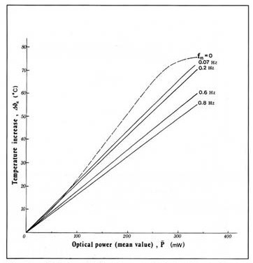
Figure 7
Variation of the increase Δϑs in the steady-state temperature of the thermal head material as a function of the mean optical power of the incident modulated laser beam, measured for various values of the modulation/repetition frequency. The curve for the unmodulated beam (dashed line) has been traced from Figure 5 for comparison purposes.
Lastly, from inspection of the curves of figure 8 it is first deduced that the amplitude and/ or the modulation depth of the temperature oscillations provoked by an iM modulated beam of constant mean power P decrease with increasing repetition rate fm, so that practically no temperature modulation subsists after a cut-otf value fc of about 0.3 Hz (curves a and b in fig. 8). By contrast, as seen from figure 7 and more clearly from curve c of figure 8, the correspondingly induced increase in the steady-state temperature of the attacked material decreases with frequency but so slightly that, even roughly, it can be considered practically constant, particularly at higher frequencies (repetition rates). The applications of these results in the medical field have been already studied experimentally in the case of a Nd:YaG laser by other researchers, which have found that the pulsing rate of the laser beam does not influence the tissue temperature profile or the nature and extent of the tissue damage (20). Nevertheless, it is worthy noting that the degree of the temperature modulation under discussion, though not particularly affecting the steadystate temperature conditions, may play a non¬-negligible complementary role in questions such as the smoothness and precision of the cutting contour, the range and extent of heat intrusion to the adjacent iayers, etc. accordingly it would be useless te try influencing these secondary effects by means of repetition rates exceeding the cut-off frequency fc above which- as seen- the temperature modulation of the tissue becomes negligible.
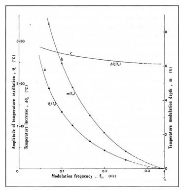
Figure 8
Variation of the amplitude ϑo of the steady-state temperature oscillation (curve a), the corresponding temperature modulation depth m (curve b), and the steady-state temperature increase (curve c) Δϑs as functions of the modulation frequency fm of the modulated laser beam, measured for a mean optical power P = 100mW.
Summary
Throughout the paper the thermal (temperature) response of a bulk thermal sensor head, such as the KTY 10 thermistor, t o a co2 laser beam has been experimentally studied with the beam, being in a cw and in an iM-modulation regime as weii. also, the corresponding influence of the (mean) optical power P and the modulation frequency (repetition rate) fm on the steadystate temperature ϑs of the targeted mass has been investigated in detail. The results of this investigation have been evaluated and interpreted with the thermal head considered as well as thermometric sensor as simulation of a living tissue.
We have thus ascertained that the application of intensity modulation, in a square-wave format, may ensure a considerable linearization of the temperature increase versus optical power curve and suppress the bothering saturation effect appearing in the case of unmodulated beam. This influence of iM modulation is accompanied by an undesired decrease of the temperature increment, but this effect is rather weak and may be considerably reduced by choosing a small or even moderate modulation frequency fm (in the present case, of the order of 0.1 Hz). in accordance to the above, the iM modulation of the beam can be employed either for improving the performance of an automatic control system commanding the beam-induced tissue temperature or for iiberating the attacked tissue from the temperature saturation drawback.
In addition, it has been found that the intensity modulation of the laser beam is transformed into a modulation (oscillation) of the correspondingly indu ced steadystate temperature only for repetition rates fm iower than a cut-of frequency fc and the so obtained temperature modulation depth m decreases when fm increases. This fact, which has been also observed when a Kr-ion laser was used by the first of the authors et al, imposes employing repetition rates clearly iower than fc whenever a strongly modulated steadystate tissue temperature is required in medical laser applications.
In conclusion, the intensity modulation of a laser beam iowers (to a small extent only) the level of the achievable temperature increase of the irradiated material but offers very useful advantages such as the enhancement of the temperature-to-optical power linearity, the suppression of saturation effects, and possibility for modulation of the steady-state temperature. obviously, direct investigations of the above in pure biological tissues would be particularly useful.
Bibliography
- Carouth JaS, McKenzie al. Medica; lasers: science and clinical practice. 2nd ed. Bristol/london: adam Hilger 1986.
- Hillenkamp f. laser radiation and tissue interaction. Health Physics, 1989, 56: 613-8.
- Mùiier o, Schaldach B. Basic laser tissue interaction. in:Berlien HP, Giamino G, Mùiier G, ringelhan H,Scaldach B, eds. advances in laser Medicin il. Berlin. (Safety and laser – Tissue lnteraction european com¬munity Medical laser concerted action Programme)1989; 17-25.
- Yoon G, welch aJ, Motamedi M, Gemert McJ Development and application of three-dimensional iight distribution model for laser irradiated tissue. ieee Journal of Quantum electronics 1987: Qe-23: 1721-33.
- Furzikov NP. Different iasers for angioplasty: thermooptical comparison. ieee Journal of Quantum eiectronics 1987; Qe-23: 1751-5
- Zweig aD, weber HP. Mechanical and thermal parameters in pulsed laser cutting of tissue ieee Journal of Quantum eiectronics 1987; Qe-23:178793
- Canestri f. Beam-tissue interaction: iong-term modeling. lasers & optronics,1990, 9: 55-7.
- Moore Kc. calderhead rG. The clinical application of low incident power density 830 nm Gaa1as diode laser radiation in the therapy of chronic intractable pain: an historical and optoelectronic rationale and clinical review. international Journal of optoelectronics, 1991, 6:503-20.
- Mùller G, Harnoss H, Karr H, Dorschel K, Berlien H-P Photoablation, a question of wavelength? in: Marshail J, ed. laser Technology in opthalmology. amsterdam: Kugler & Ghedini Pubi 1988; 221-7.
- Jacques Sl, Prahl Sa. Modeling optical and thermal distribution in tissue during laser irradiation. laser in Surg Medicine,1987, 6:497-503.
- Jones Be. lnstrumentation, measurement, and feedback. 4th, ed. New Delhi: Tata/McGraw-Hill 1986; 73-5.
- Bentley JP. Principles of measurement systems. 2nd ed. New York: longmag/J wiley & Sons 1988; 144-7
- Usher MJ Sensors and transducers. 1st ed. london: Macmillan co 1985; 81-8
- Theofanous N, raftopoulou a, Tsitomeneas S, arapoyanni a. influence of electrooptic modulation of a Krypton-iaser beam on thermal detector response. in: carabelas aa, letardi T, eds. Proceedings of the 1st Gr-i international conference on New laser Technologies and applications. Bologna: Società italiana di fisica 1988; 237-44
- Halldorson T, langerholg J. Termodynamic analysis of laser irradiation of biological tissue. applied optics, 1978; 17: 3948-58.
- laufer G. Primary and secondary damage to biological tissue induced by laser irradiation. applied optics,1983, 22:676-81.
- Darchuck JM, Migliore lr. Guidelines for laser cutting. laser & applications 1985:4 (Sept): 91-7.
- Kaplan r. YaG lasers in breakdown-mode surgery. lasers & applications 1984:3 (Nov): 53-9.
- VanGemert MJc. laser-tissue interaction. in: abstracts of the 3rd congress of the european laser Society (laser in Medicine). amsterdam, Nov 1986; 56.
- Matthewson K, coleridge-Smith P, Northfield Tc, Bown SG.comparison of continuous wave and pulsed excitation for interstitial Nd:YaG laser induced hyperthermia. in:abstracts of the 3rd congress of the european laser Society (laser in Medicine). amsterdam Nov 1986; 102.