Energy for Health, Vol. 24, 2024
G. Insero1*, G. Romano1
1 Department of Experimental and Clinical Biomedical Sciences “Mario Serio”, University of Florence Viale G. Pieraccini 6, 50139 Florence, Italy
* E-mail: [email protected]
Abstract
Laser therapies are based on the principle of light absorption by tissues and rely on three types of interactions: photochemical, photothermal and photomechanical interactions. In the first case, the photon energy is employed to trigger photochemical reactions, in the second the energy is converted into heat, while in the latter case mechanical effects are elicited. The prevalence of one or the other effect can be studied by considering the “chart of photo-induced effects” as detailed below. Generally speaking, laser interaction with tissue is governed by the following parameters: (i) laser wavelength, irradiance and light delivery mode for the laser source; (ii) the absorption and scattering properties (i. e. absorption and scattering coefficients) for the tissue. Irradiance, defined as the amount of light energy impinging on the tissue per unit of time and area, determines the intensity of the laser beam interacting with the target tissue.
After introducing a classification of the photo-induced effects, we will concentrate on the photothermal ones, analyzing their role in both therapeutic effects and safety issues.
Introduction
Chart of photo-induced effects
When laser light interacts with biological tissues, different effects can take place, depending on both the tissue’s properties and the parameters of the light source [1-3]. These effects can be initially divided into two main categories: radiative processes, which involve the emission of energy in the form of light or other electromagnetic radiation, and non-radiative processes, where the absorbed energy is dissipated through mechanisms such as heat generation or molecular vibrations, without the emission of radiation. In this discussion, we will focus solely on non-radiative processes, as radiative effects are generally negligible in the context of photo-therapies and are mainly relevant for diagnostic applications only.
Non-radiative processes can be categorized using a diagram (Fig. 1) that associates them with two critical factors: the light irradiance (E) at the tissue surface and the duration of the laser exposure (τlaser). The irradiance is generally measured in Watt / cm2 and can either be calculated from the laser emission characteristics — factoring in potential changes due to reflections, beam expansion in air, or interaction with other tissues — or measured directly with a power meter, which generally returns a power value. Then, accurately determining E requires proper estimation of the beam’s spot size at the air-tissue interface, which may involve various tools. These range from simple instruments like rulers or calipers to more advanced methods such as Knifeedge techniques. Another option involves using light-sensitive films, such as Gafchromic™ films, which can provide a resolution of up to 50 μm in the UV-visible range. However, newer versions of these films like the EBT3 model show reduced sensitivity to visible light, though they still perform well in certain UV ranges. A reasonably precise estimate of τlaser by the operator is often sufficient to predict the primary effects, as outlined in Fig. 1. This estimation is valid when τlaser corresponds directly to the treatment duration or is appropriately derived from the operational characteristics of the laser being used in the case of pulsed lasers.
Once both E and τlaser are established, their product yields the energy fluence [J/m²], which represents the energy per unit area delivered to the biological tissue. In Fig. 1, inclined lines are characterized by constant energy fluence values, aiding in the visualization of how the same energy fluence can be achieved under different combinations of E and τlaser. Based on the increasing energy fluence values, the three main photo-induced effects are identified: photochemical, photothermal, and photomechanical. It can be noticed that photochemical interaction is predominant for lower irradiance values and higher interaction time; if these two parameters increase, photothermal interactions become visible until they eventually account for the primary interaction mode. It is important to remember that more than one effect can be present at the same time; the chart in Fig. 1 can help in predicting if and which of them will be predominant in given irradiation conditions.
Photothermal Effects
When a biological tissue is irradiated with a laser beam, the photons may be absorbed by various molecules within the tissue, known as chromophores. This absorption leads to an increase in the energy state of these molecules, which then dissipate energy as heat through molecular vibrational and rotational modes. The efficiency of this process is highly dependent on the tissue’s specific optical properties, particularly its absorption coefficient, which varies with the wavelength of the incident light.
The photothermal effect is largely determined by the tissue’s endogenous absorbers, including water, hemoglobin, and melanin. These chromophores absorb light at specific wavelengths, making the photothermal response wavelength-dependent. For instance, water strongly absorbs infrared radiation, while melanin and hemoglobin absorb light primarily in the visible and near-infrared regions. This wavelength-dependent absorption must be carefully considered when selecting lasers for therapeutic purposes, as it dictates both the depth of penetration and the nature of the thermal effect produced in the tissue.
For a more detailed discussion on the absorption properties of optical radiation by the most prevalent chromophores in biological tissues, we refer to the review by Martins et al. 2023 [5]. In that study, the absorption and effective absorption coefficients μa and μeff are precisely defined, which is the physical parameter that quantifies the overall light absorption. It is important to highlight that a specific molecule absorbs light differently depending on the wavelength. Therefore, a single absorption value cannot be applied across a broad range of wavelengths. Instead, for each specific λ, the corresponding absorption coefficient must be used. Failing to do so could result in significant errors in the analysis or application. This wavelength-dependent absorption is critical in applications like laser therapy, where precise energy deposition in the tissue is required for therapeutic efficacy and to avoid unintended damage.
Understanding and applying the correct absorption coefficient for the relevant wavelength ensures that the interaction between light and tissue is accurately modeled, leading to more controlled and predictable outcomes.
For a more detailed discussion on the pathological effects of temperature increase in tissues, we refer to Douplik [1] and Thomsen [6]. For a quick overview of photothermal effects, Table 1 summarizes the main effects that can be induced depending on the temperature reached by the biological tissue. Some of these effects can be classified as reversible or irreversible, based on whether the tissue can return to its normal state once the temperature increase ceases. Reversible damage allows the cell or tissue to recover, while irreversible damage occurs rapidly, typically within seconds to minutes, and cannot be repaired through natural healing processes, which usually take days or weeks. Fig. 2 illustrates the conditions under which irreversible biological effects are achieved.
| Tissue Temperature | Photothermal Effects |
|---|---|
|
42°C – 45°C |
Protein structural changes, hydrogen bond breaking, retraction |
|
45°C – 50°C |
Enzyme inactivation, changes in membrane permeabilization, oedema |
|
50°C – 60°C |
Coagulation, protein denaturation |
|
~ 80°C |
Collagen denaturation |
|
80°C – 100°C |
Dehydration |
|
> 100 °C |
Boiling, steaming |
|
100°C – 300°C |
Vaporisation, tissue ablation |
|
> 300 °C |
Carbonisation |
Table 1
Photothermal effects of laser-tissue interaction as function of the tissue temperature.
The temperature increase in biological tissue can be induced using an optical source that operates either in continuous or pulsed mode. In the first case, with continuous operation, energy is delivered steadily and continuously until the operator decides to stop the laser emission, resulting in gradual and uniform heating of the tissue. In contrast, pulsed optical sources emit laser radiation in short, repeated bursts. The pulse duration, repetition rate, and delivered energy are parameters that depend on the specific type of source used and may or may not be adjustable by the operator. The overall treatment duration, therefore, corresponds to a different number of pulses delivered. This method generally allows heat to be concentrated in localized areas, limiting thermal diffusion to surrounding tissues and reducing the risk of collateral damage. In fact, if we consider pulsed lasers, the photothermal effects depend on the pulse duration ( tpulse) and the specific tissue thermal properties, which are represented by the so-called “thermal relaxation time” ( tthermal). This characterizes the time with which heat is dissipated in the tissue, starting from the volume where laser absorption takes place. Formally, tthermal is defined as a function of the laser penetration depth and the tissue thermal diffusion coefficient, and will be the object of a specific article. Here, we would just like to mention that the following two cases can be discussed: (i) tpulse << tthermal; (ii) tpulse >> tthermal. In case (i), heat is confined in the volume where laser absorption takes place, leading to a high and local temperature increase. Case (ii) represents the opposite occurrence, where heat is dissipated in the surrounding regions during the photothermal energy release, therefore heating a greater tissue volume but limiting the temperature increase. The choice of laser wavelengths and laser pulse duration determines the occurrence of one or the other case.
The heat generated during laser-tissue interaction can be exploited in therapy [7-10], both in the treatment of musculoskeletal pathologies and in regenerative medicine, according to the modality and timing of heat release. At the same time, photothermal effects are associated with laser-safety issues, for both the patient and the operator, in the case they are not the main desired interaction pathway when using laser light in the medical field [11]. When the energy delivered by the laser is too high, or the exposure time is too long, the resulting temperature rise can cause tissue damage, leading to burns or other injuries. To mitigate these risks, stringent safety protocols are required, encompassing both the technical parameters of laser systems and the training of personnel. Compliance with safety standards, such as those set by international guidelines, ensures that the therapeutic benefits of laser treatment are realized without compromising safety.
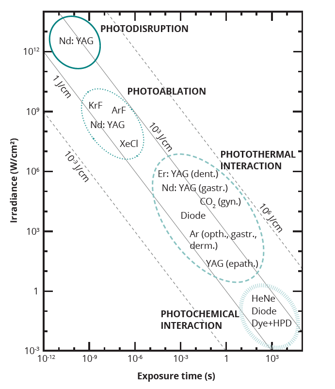
Map of photo-induced effects reported as function of the exposure time τlaser, expressed in second (s) and light source irradiance E, in W/cm2 [4].
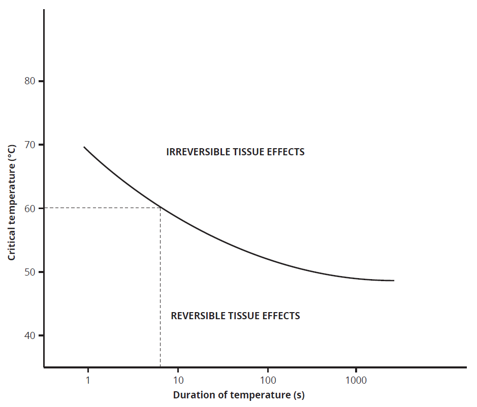
Critical temperatures for the occurrence of cell necrosis. Image from [1].
Conclusions
In conclusion, the interaction between laser light and biological tissue is a complex process governed by the wavelength-dependent absorption of light by endogenous chromophores. This interaction forms the basis for photothermal effects, which are widely utilized in medical therapies. The proper understanding and management of these effects are crucial not only for achieving therapeutic efficacy but also for adhering to laser safety protocols, ensuring that laser treatments are both effective and safe for patients and operators alike.
Bibliography
- Douplik, G. Saiko, I. Schelkanova, V. V. Tuchin, The response of tissue to laser light. In: Lasers for Medical Applications, Woodhead Publishing Series in Electronic and Optical Materials, 2013, pp 47-109.
- Tuchin. Tissue Optics: Light Scattering Methods and Instruments for Medical Diagnosis. SPIE-International Society for Optical Engine, 2007.
- H. Niemz, Laser-Tissue Interactions: Fundamentals and Applications, Springer Berlin, 2007.
- Insero, F. Fusi and G. Romano. The safe use of lasers in biomedicine: Principles of laser- matter interaction. J. Public Health Res., 2023, 12(3).
- Martins, H. F. Silva, E. N. Lazareva, N. V. Chernomyrdin, K. I. Zaytsev, L. M. Oliveira, and V. V. Tuchin. Measurement of tissue optical properties in a wide spectral range: a review. Biomedical Optics Express, 2023, 14(1): 249-298.
- Thomsen. Pathologic analysis of photothermal and photomechanical effects of laser-tissue interactions. Photochem. Photobiol. 1991, 53(6): 825-835.
- Zhi, et al. Photothermal therapy. Journal of Controlled Release, 2020, 325: 52-71.
- Yang, et al. Low temperature photothermal therapy: Advances and perspectives., Coordination Chemistry Reviews, 2022, 454: 214330.
- Yi, Q. Duan, and F. Wu. Low-temperature photothermal therapy: strategies and applications., Research, 2021, 7: 9816594.
- Liping, et al. Recent advances in selective photothermal therapy of tumor., Journal of nanobiotechnology, 2021, 19: 1-15.
- Insero, et al. Risks associated with laser radiation reflections in a healthcare environment: a surface reflectance study in the range 250 nm – 25 μm., Healthcare in Low-resource Settings, 2024.