Energy for Health, Vol. 16, 2017
S. Meggiolaro, S. Tention, G.M. De Benedictis
Meggiolaro S., Tention S.: Ambulatorio Veterinario Thermal Physiopet
De Benedictis GM: Department of Animal Medicine - Productions and Health - University
of Padova
Abstract
Degenerative Joint Disease (DJD) is one of the most common and disabling orthopaedic conditions of pets. The most recent therapeutic approach consists in the combination of different therapeutic options, such as the use of conventional drugs, the use of alternative treatments (i.e. homeopathy, phytotherapy, acupuncture), the oral administration of chondroprotectors (i.e. nutraceuticals), body weight control, rehabilitation and correct home management.
This study compared the efficacy on arthritic pain control of a physical therapy protocol, including MLS® treatment and hydrotherapy, versus traditional nonsteroidal anti-inflammatory drug (NSAID) therapy.
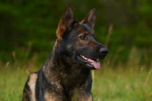
Sixteen Labrador dogs, older than 5 years and affected by osteoarthritis have been included in the study.
After the baseline visit, the animals matching inclusion criteria have been allocated to one of the treatment groups. The treatment efficacy has been assessed at 15 and 45 days via pet owner’s evaluation, using the Liverpool Osteoarthritis in Dogs (LOAD) and the Canine Brief Pain Inventory (CBPI), and by the clinical assessment of a technical expert.
In both groups, a general improvement in symptoms has been observed, confirming that both physical therapy with MLS® and drug therapy are valuable aids in the management of pain symptoms associated with degenerative joint disease.
In particular, for the treatment of ostearthrosis, when long term treatments are necessary, MLS® laser therapy is a valid alternative to pharmacological therapy, allowing for treating old dogs without worsening the condition of other compromised organs.
Introduction
Degenerative Joint Disease (DJD) is a major condition affecting especially old and/or obese dogs, those subjects presenting genetic bone abnormalities or bone conditions and active dogs that are prone to repeated microtrauma due to intense physical activity [1].
Secondary DJD caused by trauma, articular instability or osteochondral lesion is the most common [2].
DJD has a severe impact on quality of life, due to associated pain and biofunctional limitations. Pain is the main clinical symptom of OA, therefore pain management is of outmost importance in osteoarthritis (OA) treatment, allowing improvement in both physical and psychological quality of life of the subject. Currently, there is no resolutive treatment for OA and several approaches have been investigated to address pain, inflammation and progressive degeneration, which are different aspects of the disease, leading to the so-called multimodal approach [3]. This recent approach consists in the combination of different therapeutic options, combining the use of drugs with less conventional treatments, such as homeopathy, phytotherapy and acupuncture; the oral administration of chondroprotectors, i.e. nutraceuticals; dog weight control; rehabilitation and correct home management by the pet owner. The pharmacological treatment of the DJD involves the use of NSAIDs, chondroprotective drugs and other complementary medications. This study investigates the possibility of alternative therapies that may be more suitable, especially for older dogs with unpaired general health for which drug therapy may not be appropriate. Among the different physical therapy options that were taken into account, Multiwave Locked System(MLS®) laser therapy was considered the most suitable for dog DJD treatment. MLS® therapy involves the use of two different and synchronised emissions: one with continuous/frequenced mode at 808 nm wavelength, the other with pulsed mode at 905 nm. The average power of the device is 1.1 W with a peak power of pulsed emission of 25 W. MLS® has been clinically applied for the treatment of several pathologies, including shoulder pain, lumbago, carpal tunnel syndrome, etc. MLS® pulse has been extensively characterized and its effects are well documented [4-6]. In this study a protocol that used MLS® associated with hydrotherapy treatment, was compared with the traditional pharmacological approach in dog DJD. The aim of the study was the assessment of both approaches efficacy in OA treatment, based on dog owner’s feedback and the clinical examination results, and the comparison of the results of the two treatments.
Materials and Methods
During the reference period, 25 Labrador dogs were assessed, 16 of which met the study inclusion criteria and have been enrolled. The inclusion criteria were collaborative dogs of age > 5 years with a diagnosis of OA in the elbow, hip or knee, confirmed by X-ray. Animals presenting concomitant pathologies, reduced cognitive abilities or adverse reactions to NSAIDs were excluded from the study.
A series of critical aspects, such as scattered clinical conditions, breed specific characteristics, nutritional status, pain multifactor components, pet owner time availability and economical possibilities, should be considered as they may heavily impact on treatment outcome [11]. Labrador dogs have been selected as study subjects to limit breed variability. This specific breed was chosen as it is a diffuse breed with predisposition for joint pathologies, frequently affected by OA. Additionally, Labrador dogs are generally collaborative, which simplifies the physical therapy treatment, and express pain sensation without excessive misrepresentation, as it happens for example with other breeds. The selection of strict inclusion criteria represented a limitation in terms of number of eligible subjects, but it was needed to reduce the risk of confounding factors. The study took place at the rehabilitation centre Thermal Physiopet (Montegrotto Terme, IT), from September 2015 to March 2016. Dogs were allocated to two groups, respectively group A and B, groups were homogenous for age and weight. Group A underwent to physical therapy sessions including MLS® treatment and hydrotherapy. Animals that received medications from the owner were excluded from this group. Hydrotherapy has been performed in specific pools equipped with treadmills, walking speed was set at 32m/min for 3 minutes and water level was set at the superior third part of the dog femur. The exercise was repeated 4 times with 3 minute breaks in between repetition. Animals were observed during the exercise and after the hydrotherapy session: in case of fatigue, the protocol was adjusted accordingly. After the session, the animal was clinically assessed and the laser treatment was performed.
In this study, the following treatment parameters have been applied:
- Animals with pain at palpation > 4: muscle scan phase with 18 Hz frequency and fluence of 4,81 J/cm2
- Animals with pain at palpation < 4: muscle scan phase with 36 Hz frequency and fluence of 4,81 J/cm2
- Animals with flexion-extension pain > 4: the treatment had been carried out on points, covering the whole articular surface with 18 Hz frequency and fluence 3,99 J/cm2 for a total of 12,52 J/point
- Animals with flexion-extension pain < 4: the treatment had been carried out on points, covering the whole articular surface with 36 Hz frequency and fluence 3,99 J/cm2 for a total of 12,52 J/point.
In general, 100% intensity has been used. It has been reduced to 75%. for animals with dark fur,
- Each trigger point had been treated from one to four times with the following parameters:10 Hz with 25% intensity and fluency 1 J/cm2.
The physical therapy protocol was repeated three times a week for the first two weeks, two times a week for the following two weeks and once a week up to the end of the study. Group B was treated with NSAIDs using oral administration of Carprofen (Rimadyl) with a dosage of 4mg/kg once per day (SID) for the first 7 days, followed by a dosage of 2 mg/kg SID for other 7 days. For gastroprotection, omeprazole was orally administered 20 minutes before food intake with a dosage of 0,7 mg/kg SID.
Assessment were performed at day 0 (enrolment and first treatment), 15 and 45 using:
- Pet owner’s evaluation with Liverpool Osteoarthritis in Dogs (LOAD) scale and the Canine Brief Pain Inventory (CBPI) scale,
- Technical expert clinical examination, with observation of: lameness degree (score from 0- no lameness, to 4-limb is lifted and no load bearing), muscle tonicity (score from 0 – tonic limb, to 3 – severe hypotonicity), flexion-extension pain by VAS scale (evaluation based on dog behaviour reaction), pain at palpation of the main muscle groups by VAS scale, trigger point number. When more than one limb was affected, the one with the most severe condition was scored. All the visits have been performed by the same operator, blinded to the pet owner’s assessment.
During the visit at day 0, dog specific counselling was provided to the owner in terms of dietary advice and physical activity protocol. Data were analysed using the Shapiro Wilk test, data presenting a normal distribution were expressed as mean ± standard deviation, while not normally distributed data or ordinal data were expressed as median (min-max). Intraobserver variation related to evaluation during time by the pet owner and the technical expert was considered.
Group A and group B were compared using the Student t-test and two-way ANOVA.
Friedman test was used to analyzed ordinal variables or not normally distributed differences between treatment, time and their interaction.
Statistical significance was set with p<0,05.
Results
Each owner completed the LOAD and CBPI questionnaires at days 0, 15, 45. In both groups, an improvement in the animal condition was perceived by the owners, despite it did not reach statistical significance (Table I). The results obtained in group A and B in terms of owner’s questionnaire were compared and show no statistical difference (Table II).
Lameness degree was assessed by the technical observer and scored 2,625 ± 0,91 at T0, 1,75 ± 1,03 at T15 and 1,75 ± 0,89 at T45 for Group A, while Group B obtained the following scores: 2,625 ± 0,92 at T0, 1,61 ± 1,19 at T15 and 1,87 ± 1,46 at T45. The degree of lameness decreased in both groups reaching statistical significance respect to baseline, while no difference was observed between the groups (Figure 1).
Flexion-extension pain by VAS scale (Figure 2) statistically improved during time in Group A from 6,37 ± 2,2 at T0 to 5,25 ± 1,89 at T15 to reach the score of 3,62 ± 0,74 at T45. Group B showed VAS value at T0 (6,87 ± 1,72), T15(5,25 ± 1,67) and T45(5,5 ± 1,77), that do not indicate significant modifications from baseline.
Pain at palpation statistically improved for both groups from T0 to T15 (Figure 3). The following scores were assigned respectively to Group A and B: 6,5 ± 1,77 at T0; 5,37 ± 2,13 at T15 and 4,62 ± 1,60 at T45 and 6,37 ± 1,59 at T0; 4,5 ± 1,85 atT15 and 5 ± 2,14 at T45.
Muscle tonicity (Figure 4) improved for Group A (from 1,75 ± 0,71 at T0 to 1,25 ± 1,03 at T15, up to 0,37 ± 0,74 at T45), while no changes were observed for Group B (scored 1,25 ± 1,03 at T0, 1,25 ± 0,99 at T15 and 1,26 ± 0,99 at T45).
Trigger point number (Figure 5) statistically decreased in Group A (5,87 ± 2,17 at T0; 3,75 ± 1,67 at T15 and 1,75 ± 1,39 at T45) while no change was detected in Group B(5,5 ± 2,72 at T0; 4,87 ± 2,64 at T15 and 5,15 ± 2,99 at T45).
Discussion
Pain management in OA is a key point for pet quality of life improvement and pet owners are particularly sensitive to this topic. Recently, pain management protocols have benefited from scientific research progress and allow for a better subject treatment. The current concept in OA treatment involves the combination of pain control, which is the main clinical symptom, with strategies that specifically target the degenerative, inflammatory and oxidative processes involved in OA progression. The so-called multimodal approach is the association of pharmacological treatments, such as the use of ant-inflammatory drugs, and nonpharmacological treatments, involving the use of chondroprotective agents and nutraceuticals, diet control, physical therapy and exercise. The present study aimed at comparing the effects of two different therapeutic approaches: physical therapy versus pharmacological treatment. MLS® therapy has been selected as treatment of choice, due to its large use in the veterinary centre which conducted the study and to practical advantages respect to other physical therapies, such as the fact that laser application does not require trichotomy, a procedure which is not appreciated by dog owners. OA affected dogs generally present impaired mobility, which in turn results in loss of muscular tone. To limit this, MLS® therapy has been associated to hydrotherapy, which allows muscular work, minimizing load bearing [7 -10].
The pharmacologically treated group received Carprofen, one of the most commonly used drugs in the treatment of pain associated to chronic orthopaedic conditions. Carprofen is a COX2 preferential inhibitor and thus allows OA inflammation and pain control with less than 1% side effects [3].
Another key point for the study was the pet owner’s contribution, both in terms of home management of the dog, based on the instruction received during the inclusion visit, and in terms of his evaluation of the treatment outcome assessed using the Liverpool Osteoarthritis in Dogs (LOAD) and the Canine Brief Pain Inventory (CBPI). LOAD questionnaire is more focused on animal motility, while the CBPI questionnaire focuses on pain, therefore they appear to be complementary and were used together to assess the overall health status of the animal. A limitation in the use of these questionnaires is the subjectivity related to the owner sensitivity and his emotional relationship with his dog, that may alter the perception of the real health condition of the animal [12]. To balance these factors, a clinical examination by a trained expert had been included in the study and, in fact, in many occasions this clinical assessment did not correspond to the owner’s evaluation. This can be explained by the fact that the clinical examination considered the OA grade of the dog, while the owner was likely influenced in his assessment by the knowledge of the health status of his dog before OA onset.
Another study limitation is related to the need for interpretation of the subject algic response. In relation to this, the evaluation of subjects belonging to the Group A was easier respect to the evaluation of the Group B subjects. Since the physical protocol implied longer treatments, this means that the operator spent more time and got to know more closely Group A dogs compared to Group B dogs, which have been in contact with the operator only during follow up visits. The study results show improvement in symptomatology in both groups.
This is an important achievement, and confirms that both physical and pharmacological therapies are suitable tools for OA pain management.
In our study, no significant differences have been observed between the two treatment outcomes, encouraging to consider the option of prescribing one treatment or the other based on specific animal conditions and characteristics. For instance, when there are restrictions to NSAIDs usage due to dog general health, laser therapy should be preferred as it demonstrated the same pain control and anti-inflammatory effects as drug therapy but without its side effects. To conduct a successful physical therapy, dog and owner collaboration is essential.
An interesting point that emerged from our study is the advantage of the use of laser therapy over drug therapy for treating trigger points, as only laser allows to exert a local pain relief action.
Due to the limited sample size and follow up duration, this study represents a preliminary investigation and further studies are needed to assess the most appropriate therapeutic approach to pet OA.
Conclusions
Osteoarthritis is a degenerative condition for which long term animal treatment is required. Any clinical improvement in the pet quality of life should be considered as a relevant achievement. The best clinical approach to articular degeneration involves a multimodal and personalised management of the disease and of the related pain, combining traditional and more innovative treatments, and requires the owner to play an active role in home management.
In conclusion, the results of this study demonstrate that both physical therapy and pharmacological therapy are able to improve the general clinical conditions of OA affected dogs. Physical therapy allows to treat with no side effects even old and compromised animals and can be proposed as a valid alternative to traditional pharmacological therapy.
Acknowledgements
The Authors would like to thank Elena Tognato for her support and ASA S.r.l. for the help in defining the protocol setting for the laser treatment.
References
- Loeser R.F., Goldring S.R., Scanzello C.R., Goldring M.B. Osteoarthritis. A disease of the joint as an organ. Arthritis and Rheum, 2012, 64(6): 1697-1707.
- Beale B.S., Goring RL. Malattia articolare degenerativa (artrosi). In: Bojrab MJ, eds. Le basi patogenetiche delle malattie chirurgiche nei piccoli animali. Giraldi, 2001, pp 974-985.
- Zizzadoro C., Belloli C. Farmaci per il controllo dell’infiammazione. In: Carli S., Ormas P., Re G., eds. Farmacologia veterinaria. Idelson-Gnocchi, 2009, pp 455-489.
- Monici M., Cialdai I., Romano G., Corsetto P.A., Rizzo A.M., Caselli A., Ranaldi F. Effects of IR laser on myoblasts: prospects of application for counteracting microgravity-induced muscle atrophy. Microgravity Science and Technology, 2012, 25(1): 35-
42. - Monici M., Cialdai F., Ranaldi F., Paoli P., Boscaro F., Moneti G., Caselli A. Effect of IR laser on myoblasts: a proteomic study. Mol Biosyst, 2013, 9(6):1147-1161.
- Assis L., Moretti A., Parizotto N.A. Low level Laser therapy (808 nm) contributes to muscle regeneration and prevents fibrosis in rat tibialis muscle after cryolesion. Lasers Med Sci, 2013, 28(3): 947-955.
- Levine D., Tragauler V., Millis D.L. Percentage of normal weight bearing during partial immersion at various depth in dogs. Proceedings 2nd International Symposium on Rhehabilitation and Physical Therapy in Veterinary Medicine, Knoxville, Tenn., 2002, 189-190.
- Millis D., Levine D. The role of exercise and physical modalities in treatment of osteoarthrosis. Vet Clin North Am Small Anim Pract, 1997, 27(4): 913-930.
- Downer A.H. Whirlpool therapy for animals, Mod Vet Pract,1977, 58(1): 39-42.
- Dragone L. Idroterapia. In: Dragone L., eds. Fisioterapia riabilitativa del cane e del gatto. Elsevier Masson, 2011, pp 57-75.
- Fox S.M., Millis D. Gestione multimodale dell’artrosi del cane. INNOVET-EV, Italia, 2011.
- Fonda D. Dolore e analgesia negli animali. Le Point Veterinarie Italie, Milano, 2009