Energy for Health, Vol. 25, 2025
Sisti Valentina Maria, DVM
Responsible for Veterinary Rehabilitation, University Veterinary Teaching Hospital, Matelica, Italy
Abstract
Pulsed electromagnetic fields (PEMFs) are increasingly applied in veterinary rehabilitation as non-invasive and safe adjuncts, but clinical casebased evidence remains limited. This article describes three canine patients in which PEMF was integrated into multimodal rehabilitation protocols. Scooter, a 16-year-old Jack Russell Terrier with suspected cervical spinal cord contusion, underwent a two-phase PEMF protocol and regained autonomous ambulation with near-complete cervical range of motion. Yago, an 8-yearold Labrador Retriever with acute non-compressive nucleus pulposus extrusion at C4–C5, improved from non-ambulatory tetraparesis to autonomous gait within one month, with only mild residual deficits after PEMF therapy during hospitalization and follow-up. Zoe, an adult mixedbreed female surgically treated for cranial cruciate ligament rupture, received seven PEMF sessions and showed increased quadriceps circumference and reduction of lameness from grade IV to grade I. All dogs tolerated PEMF well, and consistent functional improvements were observed. These cases suggest that PEMF is a safe and adaptable modality that may support neurological and orthopedic recovery in dogs, warranting further controlled studies to confirm efficacy and optimize treatment parameters.
Introduction
Pulsed electromagnetic fields (PEMFs) are increasingly employed in veterinary rehabilitation as safe, non-invasive adjuncts for neurological and orthopedic disorders. Their therapeutic rationale is supported by a solid body of in vitro and preclinical studies. At the cellular level, PEMFs enhance fibroblast proliferation and cytoskeletal remodeling while reducing pro-inflammatory cytokine release [1,2]. They also promote myoblast differentiation [3] and increase Schwann cell proliferation without altering phenotype [4], supporting both muscle regeneration and neuroprotection. Moreover, PEMFs activate repair-related intracellular pathways such as MAPKs [5].
Preclinical canine studies have shown accelerated bone healing after osteotomy [6] and improved cartilage integration with tissue-engineered constructs [7]. Clinically, PEMF has been associated with reduced pain and improved mobility in dogs with osteoarthritis [8] and functional recovery in severe traumatic or neurological cases [9]. In human medicine, meta-analyses confirm beneficial effects on function in osteoarthritis [10] and improvements in pain and disability in chronic low back pain [11].
Despite this growing evidence, veterinary clinical reports remain limited, particularly regarding PEMF use as part of multimodal rehabilitation. This article presents three canine cases—two neurological and one orthopedic—in which PEMF was integrated into rehabilitation protocols. The aim is to describe clinical outcomes and relate them to current biological and translational evidence.
Materials and Methods
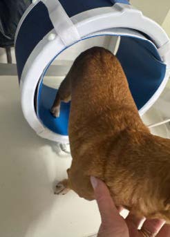
Treatments were carried out using the PMT QS device (ASA S.r.l, Arcugnano, Italy). The system allows the application of PEMFs through different applicators (Flexa and Cylinder), with adjustable parameters for intensity, frequency, magnetic flux density and treatment duration. Therapy was integrated into multimodal rehabilitation, including passive manual therapy, proprioceptive training and, when required, neuromuscular electrostimulation. Protocols lasted between four and eight weeks and were tailored to each case.
Case Presentations
Case 1 – Scooter
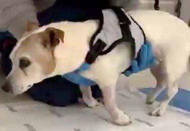
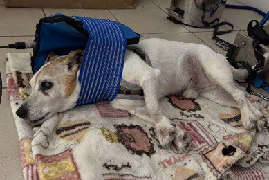
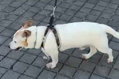
Scooter, a 16-year-old male Jack Russell Terrier, was admitted after a fall, arriving in cardiopulmonary arrest. He was resuscitated and stabilized, and clinical suspicion was of a cervical spinal cord contusion. At discharge, he exhibited non-ambulatory tetraparesis, more severe in the thoracic limbs. Rehabilitation was initiated with passive mobilizations, proprioceptive training, and pulsed electromagnetic fields (PEMF).
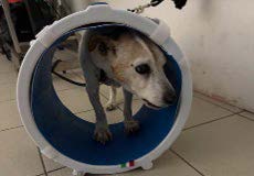
PEMF therapy followed two individualized protocols that were adjusted empirically according to Scooter’s tolerance and neurological progress. The Flexa applicator was first employed in recumbency, and the Cylinder was later adopted as patient’s condition stabilized. Details of the applied parameters are provided in Table 1. The patient was cooperative, which facilitated the protocol. After ten sessions, Scooter regained autonomous ambulation, cervical range of motion was nearly complete, and proprioception was restored, although mild thoracic limb deficits remained.
Case 2 – Yago
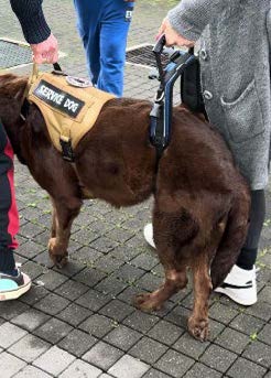
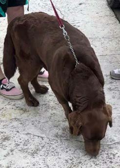
Yago, an 8-year-old male Labrador Retriever (43 kg), presented with acute tetraparesis following sudden vocalization. MRI confirmed an acute non-compressive nucleus pulposus extrusion (ANNPE) at C4–C5 with focal endomedullary myelopathy.
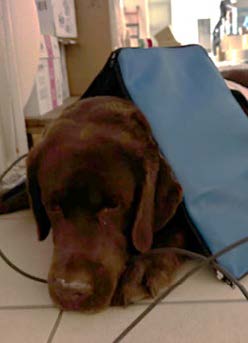
At presentation, he showed flaccid paraparesis, thoracic limb weakness, and cervical pain. Rehabilitation included pharmacological management, manual therapy, proprioceptive exercises, electrostimulation, and PEMF. During hospitalization, PEMF therapy was delivered using the Cylinder applicator through two sequential protocols. After discharge, sessions were performed weekly using the same settings, and subsequently at longer intervals, with the choice of applicator and parameters adjusted empirically according to clinical progress. Detailed parameters are reported in Table 1.
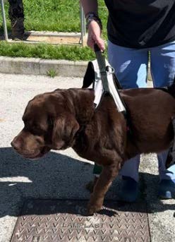
Although temperamentally lethargic, Yago tolerated the sessions well and allowed full completion of the rehabilitation program. By discharge, cervical pain and muscle tone had improved. At one month, he regained autonomous ambulation, with mild residual paraparesis and intermittent urinary incontinence. By two months, recovery had stabilized, and maintenance PEMF sessions were continued to support functional recovery.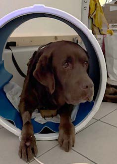
Case 3 – Zoe
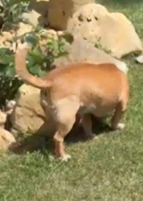
Zoe, an adult mixed-breed female, presented with grade IV lameness of the right hindlimb after trauma. Orthopedic examination confirmed rupture of the cranial cruciate ligament, surgically stabilized with an extracapsular technique. Rehabilitation began two days post-surgery and included electrostimulation and PEMF.
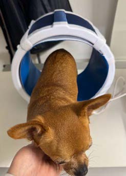
Because she tended to sit on the unaffected limb when positioned on the Flexa, PEMF was applied exclusively with the Cylinder. Each session consisted of three sequential protocols with varying frequencies (see Table 1).
Across seven sessions over six weeks, Zoe’s quadriceps circumference increased by 1.2 cm, lameness improved from grade IV to grade I, and gait became autonomous. Therapy was discontinued prematurely for economic reasons, yet significant functional recovery had been achieved.
Results
All three dogs completed the planned PEMF rehabilitation protocols without adverse effects, and overall compliance was good. The parameters applied, session schedules and applicators used are summarized in Table 1, which illustrates the progressive adaptation of treatment to each clinical condition. Clinically, consistent improvements were observed. Scooter regained autonomous ambulation with near-complete cervical range of motion, though mild deficits persisted in the thoracic limbs. Yago improved from acute non-ambulatory tetraparesis to autonomous gait within one month, with only mild residual paraparesis and intermittent urinary incontinence. Zoe recovered muscle mass and joint stability, with lameness improving from grade IV to grade I and restoration of autonomous gait, despite premature interruption of the program.
Across all cases, PEMF therapy appeared safe, well tolerated, and associated with meaningful neurological or orthopedic recovery when integrated into multimodal rehabilitation protocols.| Case | Phase | Weeks | Frequency (N° Sessions) | Applicator | Protocol | Intensity (%) | Frequency (HZ) | Field Strength (G) | Duration (Min) |
|---|---|---|---|---|---|---|---|---|---|
|
Scooter |
A |
Week 1 |
Daily
(5 sessions) |
Flexa |
1 + 2 |
85 + 85 |
10 + 100 |
32 + 17 |
30 + 10 |
|
B |
Week 2 |
Alternate days
(3 sessions) |
Flexa |
1 + 2 |
85 + 85 |
10 + 100 |
32 + 17 |
30 + 10 |
|
|
C |
Week 3 |
Every two days
(2 sessions) |
Cylinder |
3 |
70 |
5 |
50 |
35 |
|
|
Yago |
A |
Week 1 |
Daily
(3 sessions) |
Cylinder |
1 + 2 |
100 + 50 |
50 + 100 |
60 + 20 |
35 + 15 |
|
B |
Weeks 2-4 |
Weekly
(3 sessions) |
Cylinder |
1 + 2 |
100 + 50 |
50 + 100 |
60 + 20 |
35 + 15 |
|
|
C |
Weeks 5-7 |
Weekly
(3 sessions) |
Flexa |
3 |
90 |
25 |
37 |
45 |
|
|
D |
Weeks 8-11 |
Every ten days
(3 sessions) |
Flexa |
3 |
90 |
25 |
37 |
45 |
|
|
Zoe |
A |
Weeks 1-2 |
Twice weekly
(4 sessions) |
Cylinder |
1 + 2 + 3 |
65 + 50 + 60 |
50 + 100 + 5 |
35 + 23 + 40 |
25 + 15 + 15 |
|
B |
Weeks 3-4 |
Weekly
(2 sessions) |
Cylinder |
1 + 2 + 3 |
65 + 50 + 60 |
50 + 100 + 5 |
35 + 23 + 40 |
25 + 15 + 15 |
|
|
C |
Week 6 |
After 15 days (1
session) |
Cylinder |
1 + 2 + 3 |
65 + 50 + 60 |
50 + 100 + 5 |
35 + 23 + 40 |
25 + 15 + 15 |
Table 1
Detailed PEMF rehabilitation protocols applied to the three canine cases.
Discussion
The three cases described illustrate the role of pulsed electromagnetic fields (PEMFs) as supportive tools in multimodal rehabilitation of both neurological and orthopedic conditions in dogs. Despite differences in etiology and severity, PEMF was consistently associated with functional recovery, reinforcing its clinical value as a flexible and well-tolerated adjunct.
A key insight emerging from these cases is the importance of tailoring PEMF protocols to each patient’s specific clinical conditions and therapeutic response. In the first two cases, the choice between the Flexa applicator and the Cylinder was guided by the progression and localization of the pathology. In contrast, Zoe was treated exclusively with the Cylinder, as the animal’s positioning in the lateral recumbency on the healthy side prevented the magnetic field of the Flexa from adequately reaching the operated limb. The Cylinder, by generating a uniform magnetic field throughout its interior, ensured consistent exposure to the target area. This adaptability highlights how PEMF therapy can be pragmatically integrated into multimodal rehabilitation program, effectively complementing manual therapy, proprioceptive training and electrostimulation.
The outcomes observed are consistent with mechanistic studies showing that PEMFs promote fibroblast activity and reduce inflammatory mediator release [1,2], stimulate myoblast differentiation [3], and support Schwann cell proliferation [4]. These cellular effects translate into improved tissue repair and neuroprotection, in line with our neurological cases. Clinically, Kimram et al. [9] reported recovery in a polytraumatized dog treated with PEMF, while Sisti et al. [8] demonstrated significant pain reduction and improved mobility in dogs with osteoarthritis, paralleling the improvements seen in Zoe. Preclinical canine models have also shown accelerated bone healing [6] and enhanced cartilage integration [7], supporting the orthopedic rationale. The translational relevance is further reinforced by human studies. A meta-analysis in knee osteoarthritis reported functional improvement with PEMF even when analgesic effects were inconsistent [10], while a systematic review in chronic low back pain confirmed significant reductions in pain and disability [11]. These findings suggest that functional benefits may represent the most robust clinical outcome of PEMF therapy across species.
Practical aspects also deserve consideration. All patients tolerated the therapy without adverse effects, and owner compliance was crucial in maintaining continuity. In Scooter and Yago, highly motivated owners supported prolonged treatment, while in Zoe economic limitations led to early discontinuation despite evident benefits. These real-world factors underline the importance of integrating PEMF into feasible, individualized protocols rather than rigid treatment schemes.
Taken together, these observations strengthen the view of PEMF as a safe, adaptable and biologically plausible adjunct in veterinary rehabilitation. Although improvements cannot be attributed to PEMF alone due to the multimodal approach, the convergence of positive outcomes across different pathologies, combined with supporting experimental and clinical evidence, provides a strong rationale for its use. Controlled studies remain necessary to better define optimal treatment parameters and indications.Conclusions
In this series of three canine cases, pulsed electromagnetic field therapy was integrated into multimodal rehabilitation protocols and was consistently safe, well tolerated and associated with meaningful clinical improvements. Dogs affected by both neurological disorders (cervical contusion, ANNPE) and orthopedic condition (post-surgical cruciate ligament rupture) regained ambulation, improved proprioception, muscle trophism, with clear functional benefits despite differences in age, severity and treatment duration.
These observations reinforce the concept that PEMFs represent a versatile adjunct in veterinary physiotherapy, applicable across neurological and orthopedic domains. The ability to adapt protocols to the clinical condition and to patient compliance further highlights their practicality in daily practice.
While outcomes cannot be ascribed to PEMF alone due to the multimodal setting, the consistency of results, together with mechanistic plausibility and corroborating evidence from controlled studies, provides a solid rationale for its use. Further prospective trials are warranted to optimize parameters and establish clearer clinical guidelines for veterinary application.Bibliography
- Gómez-Ochoa P, Aragón CM, Aragona P, Benedetti A, Monici M. In vitro evaluation of anti-inflammatory effects of pulsed electromagnetic fields on human fibroblasts. Energy Health, 2011, 7: 18–24.
- Sereni-Monici D, Monici M, Falsini S, Bani D. Biological effects of pulsed electromagnetic fields on fibroblast models. Energy Health, 2018, 17: 4–12.
- Sereni-Monici D, Monici M, Formigli L. Effects of pulsed electromagnetic fields on myoblast differentiation. Energy Health, 2013, 9: 12–17.
- Colciago A, Mantegazza P, Senesi P, Malacrida A, Luongo C, Monici M. Effects of PEMF exposure on survival, proliferation and phenotype of rat Schwann cells. Bioelectromagnetics, 2019, 40(5): 353–365.
- El-Makakey AA, El-Masry SM, El-Mahdy HA. Comparative biological effects of low-level laser therapy and pulsed electromagnetic field on mitogen-activated protein kinases. Energy Health, 2017, 15: 4–12.
- Inoue N, Ohnishi I, Chen D, Deitz LW, Schwardt JD, Chao EYS. Effect of pulsed electromagnetic fields (PEMF) on late-phase osteotomy gap healing in a canine tibial model. J Orthop Res, 2002, 20(5): 1106–1114.
- Stefani RM, Barbosa S, Tan AR, Setti S, Stoker AM, Ateshian GA, Cadossi R, Vunjak-Novakovic G, Aaron RK, Cook JL, Bulinski JC, Hung CT. Pulsed electromagnetic fields promote repair of focal articular cartilage defects with engineered osteochondral constructs. Biotechnol Bioeng, 2020, 117(5): 1584–1596.
- Sisti E, Vannucchi G, Monici M. Pulsed electromagnetic fields for osteoarthritis treatment in dogs: Effects on pain, range of motion and quality of life. Energy Health, 2023, 24: 22–29.
- Kimram K, Plongthong N, Promsatit W, Kaewduangjai N, Asawawongsawat P. Effects of electromagnetic therapy in a multiple traumatic dog: A case report. Energy Health, 2024, 24: 18–21.
- Chen W, Xie J, Xing D, et al. Effectiveness of pulsed electromagnetic fields on pain, stiffness, and physical function in patients with knee osteoarthritis: A meta-analysis of randomized controlled trials. Pain Res Manag, 2019, Article ID 5709308.
- Mansourian M, Vafaee R, Ghaffarpasand F, et al. Effect of pulsed electromagnetic field on pain and disability in patients with low back pain: A systematic review and meta- analysis. Biomed Res Int, 2021, Article ID 6647497.