MLS® Laser Therapy Case Study
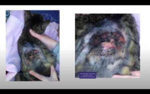
Boss, Cat Presenting Ulcer on Eosinophilic Granuloma
Species: Cat
Breed: European
Gender: Male
Age: 2 Years
Name: Boss

Clinical Report
Boss was brought into the clinic due to the presence of ulcerated, crusted plaques on his back (suspected eosinophilic granuloma) and presented significant itching. Approximately two months earlier, Boss had been bitten on the same area and developed an abscess, which, despite treatment, had not completely healed. A crust of approx. two cm2 remained in the area prior to the appearance of the ulcerated plaques. Throughout the year, the cat has been given prophylactic treatment against parasites and eats hypo-allergenic food.
Collateral Examination and Treatment Plan
A haemato-biochemical examination was carried out and found to be normal, with FIV-FeLV test being negative.
The area in question was shaved and cleansed using chlorhexidine solution, and systemic treatment using an antibiotic – clindamycin – for 5 days, with an injection of prednisone at an anti-inflammatory dosage, was administered.
In view of the cat’s temperament, a cone (recommended) could not be applied and a surgical body suit was used to protect the lesion.
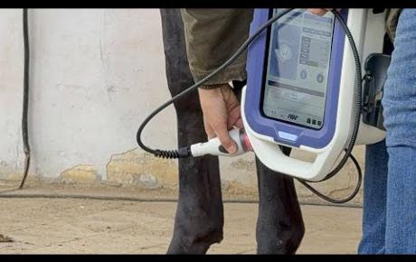
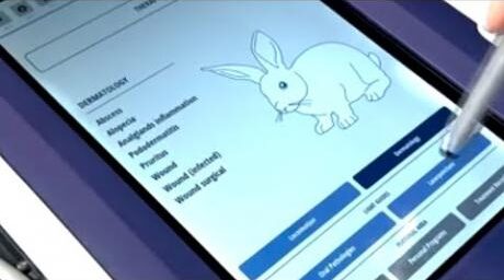
MLS® Laser Therapy was performed using the M-VET device : 8 sessions using the “Eosinophilic granuloma” protocol.
MLS® Laser Therapy & M-VET
The first seven sessions were carried out in scan mode, setting up the area to be treated. As treatment continued, the size of the area decreased from an initial 40 cm2 to 25 for the fourth and fifth treatments, then 10 for the sixth and seventh. The final application was carried out using point-by-point mode, with a single point directly on the wound.
Courtesy of Dr Irene Zanco, Montecchio Veterinary Centre – Vicenza, Italy


